
“Unveiling Hidden Anatomy: A Rare Six-Canal Mandibular First Molar”
 Dr.Razan Othman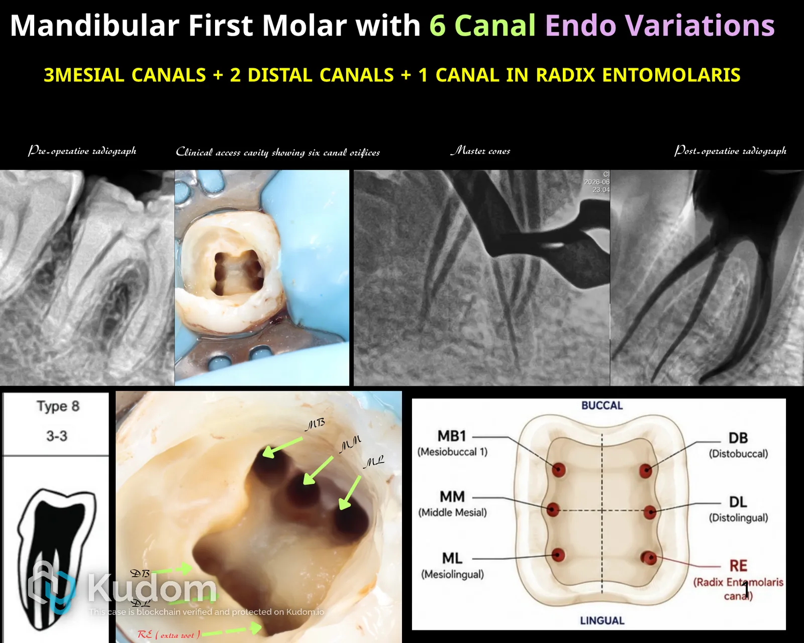
Introduction Root canal morphology of mandibular first molars can demonstrate significant anatomical variations and complex canal configurations. Understanding these anatomical differences is essential, as failure to identify additional canals remains one of the major causes of endodontic treatment failure. This case presents a rare anatomical configuration of a mandibular first molar exhibiting six distinct canals: mesiobuccal, middle mesial, mesiolingual, distobuccal, distolingual, and an additional canal within a separate Radix Entomolaris root. Careful exploration, magnification, and anatomical awareness were crucial for the successful identification, preparation, and obturation of the complete canal system. Reference Vertucci TH. Root Canal Anatomy of the Human Permanent Teeth. Oral Surgery, Oral Medicine, Oral Pathology. 1984;58(5):589–599. Diagnosis A mandibular first molar was indicated for root canal treatment.…
Leggi il caso completo — gratis
Crea un account Kudom gratuito per leggere il caso completo e sfogliare la libreria clinica verificata.