Management of a broken file during retreatment of an upper molar
 Jacopo Gotti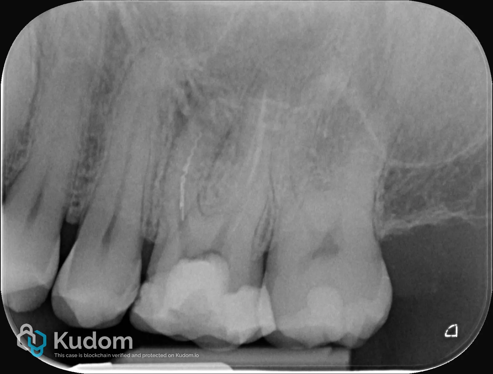
The patient was referred by a colleague for the management of a separated instrument located in the mesiobuccal root, associated with a periapical radiolucency. The patient was asymptomatic at presentation. Pre Operative Radiography The initial strategy was to remove the separated instrument due to its coronal position and easy access. However, after one session, retrieval of the fragment was unsuccessful. Therefore, the treatment strategy was modified: shaping of the mesiobuccal second canal (MB2) was performed using Procodile files, and a canal confluence was identified. Given the inability to retrieve the fragment and the presence of canal confluence, the treatment strategy focused on adequate disinfection and three-dimensional obturation of both canals while maintaining the separated instrument in situ. Irrigation was performed…
Leggi il caso completo — gratis
Crea un account Kudom gratuito per leggere il caso completo e sfogliare la libreria clinica verificata.