
Primary endodontic treatment of 3.5
 David Azaria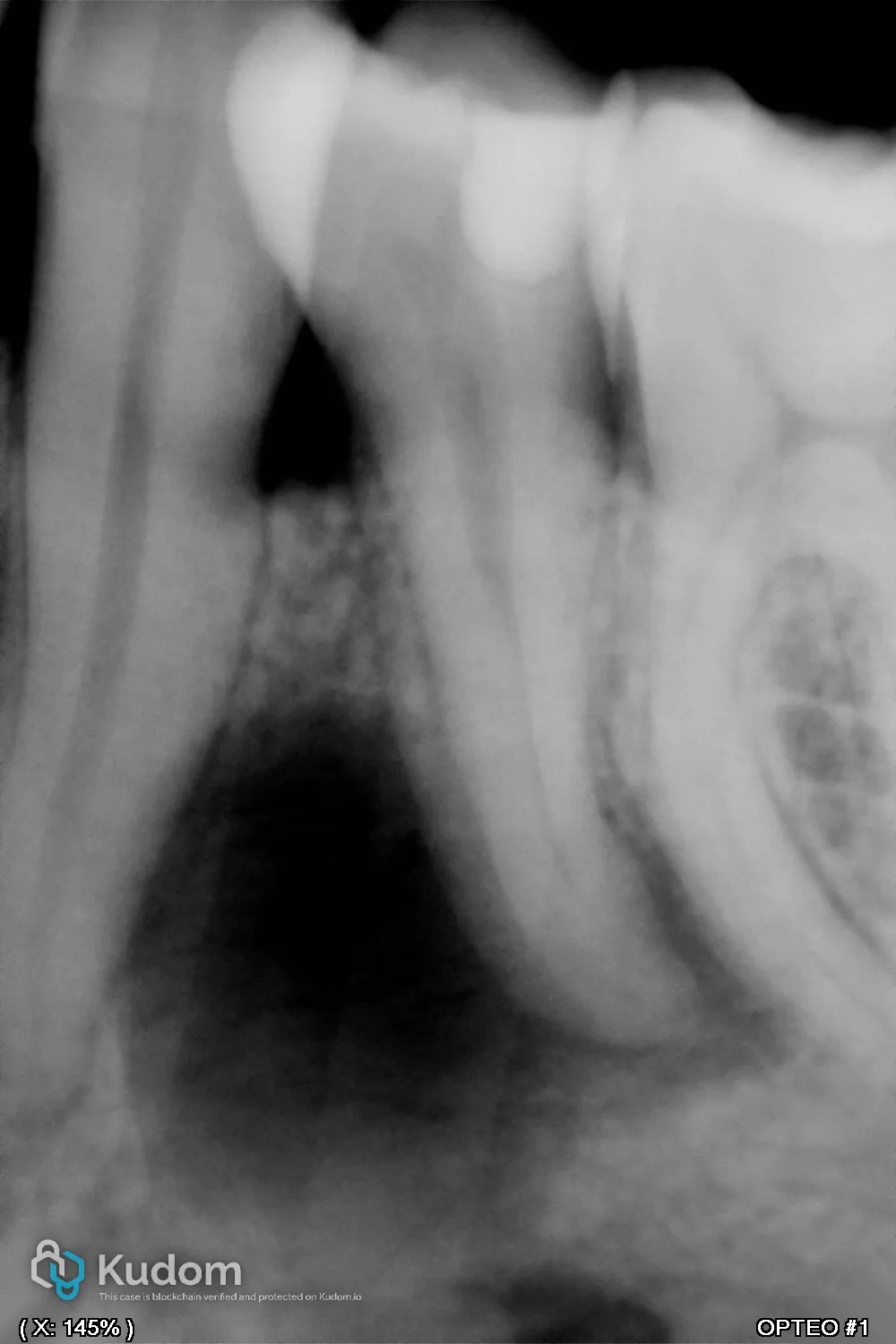
A 36-year-old male (ASA I) was referred after an incidental finding of a large apical lesion on a panoramic radiograph. Tooth 35 presented with a deep occluso-distal composite restoration, with no secondary caries or visible leakage. Clinical examination showed a negative thermal test, negative palpation, and moderately positive percussion. Radiographs revealed a periapical inflammatory lesion of endodontic origin, without resorption or sinus tract. CBCT evaluation demonstrated an uncommon and complex canal anatomy: a common coronal trunk evolving into a C-shaped canal in the middle third, and separating into two distinct apical canals (Vertucci Type V, C1 configuration). The lesion measured 9 × 13 mm and was classified as CBCT-PAI 5+D, indicating cortical bone destruction. Overall case difficulty was considered high…
Read the full case — free
Create a free Kudom account to read this case in full and browse the verified clinical library.