
Minimally Invasive Shaping of a Maxillary Second Molar with Extreme Anatomy and Trismus Using the Chameleon Dual Shaper
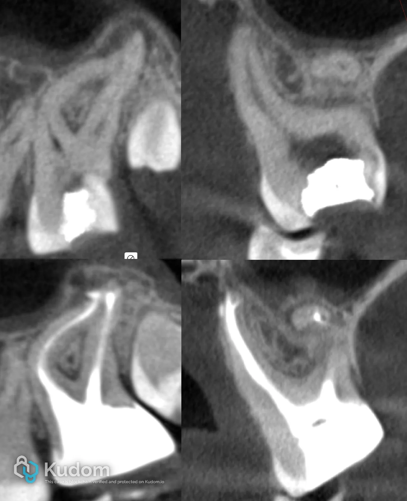
Patient and Diagnosis Patient: 29 -year-old female Chief Complaint: None (asymptomatic) Radiographic Findings: Periapical radiolucencies associated with the upper left second molar (UL7) Diagnosis: Pulp necrosis with asymptomatic apical periodontitis (UL7) Pre-operative Assessment & Challenges: Treatment Procedure: Fig.1 A pre-operative (top) and post-operative (bottom) small field CBCT. A limited field-of-view CBCT revealed three major complicating factors: Extreme multiplanar curvature of the mesiobuccal canals (MB1 and MB2) Root lengths exceeding 25 mm Severe buccal inclination of the clinical crown combined with restricted mouth opening (~25 mm), consistent with trismus These factors significantly increased the risk of procedural errors, instrument separation, and canal transportation. Treatment Strategy Access and Canal Negotiation Fig.2 An initial view of the access cavity. A conservative access cavity…
Read the full case — free
Create a free Kudom account to read this case in full and browse the verified clinical library.