Invasive External Cervical Resorption in a 40-Year-Old: Treat or Follow?
 Riccardo Tonini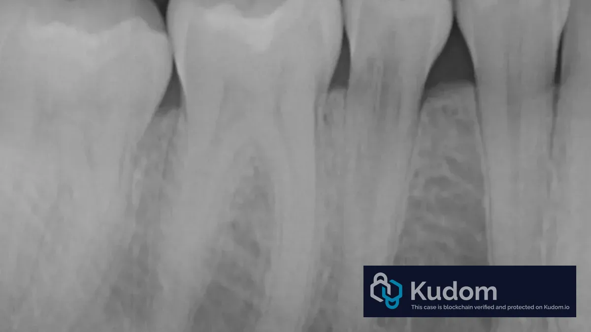
Clinical Presentation A 40-year-old patient, systemically healthy, was referred for evaluation of an incidentally detected radiolucency at the cervical third of a posterior tooth. The patient was asymptomatic and reported no history of trauma, prior orthodontic treatment, or bleaching procedures. Clinical examination revealed an intact periodontium with no signs of swelling, sinus tract, or pathological probing depths. Pulp sensitivity testing was within normal limits. Radiographic Findings Pre-operative periapical radiograph. Note the radiolucent defect at the cervical level, with apparent bone-like density partially filling the lesion. The periapical radiograph reveals a well-delineated radiolucent area at the cemento-enamel junction (CEJ). Critically, the lesion shows evidence of fibro-osseous replacement tissue partially occupying the defect — a hallmark of invasive external cervical resorption (IECR).…
Read the full case — free
Create a free Kudom account to read this case in full and browse the verified clinical library.