Endodontic Retreatment of 3.6 with large apical lesion + 11 months follow-up
 Diana Florina Chifar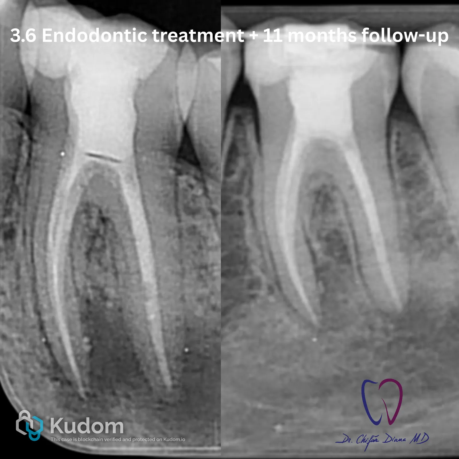
https://youtube.com/shorts/jWZ5UOYqKyE?feature=share Referred Case: Tooth 3.6 – Pulp Necrosis + Symptomatic Apical Periodontitis This patient was referred to the endodontic office after the general dentist identified a large periapical lesion on tooth 3.6. Clinical testing revealed a negative response to cold and positive axial percussion, while the pre-operative radiography showed a significant radiolucency in the periapical area — leading to a diagnosis of Pulp Necrosis combined with Symptomatic Apical Periodontitis and the need for endodontic treatment. At first glance, the tooth appeared clinically sound: no discoloration, a relatively recent direct restoration, and no visible signs of marginal infiltration or secondary caries. However, something was wrong. Upon removal of the existing restoration, a small, previously exposed pulp chamber opening was discovered —…
Read the full case — free
Create a free Kudom account to read this case in full and browse the verified clinical library.